Macular Degeneration – What is it and how is it detected?
What is macular degeneration?
Macular degeneration is a deterioration or breakdown of the macula. The macula is a small area in the retina at the back of the eye that allows you to see fine details clearly and perform activities such as reading and driving.

Central vision can be significantly affected with macular degeneration.
Although macular degeneration reduces vision in the central part of the retina, it usually does not affect the eye’s side, or peripheral, vision. For example, you could see the outline of a clock but not be able to tell what time it is.
Macular degeneration alone does not result in total blindness. Even in more advanced cases, people continue to have some useful vision and are often able to take care of themselves. In many cases, macular degeneration’s impact on your vision can be minimal.
What causes macular degeneration?
Many older people develop macular degeneration as part of the body’s natural aging process. There are different kinds of macular problems, but the most common is age-related macular degeneration (AMD). Exactly why it develops is not known, and no treatment has been uniformly effective.
Macular degeneration is the leading cause of severe vision loss in Caucasians over 65. The two most common types of AMD are “dry” (atrophic) and “wet” (exudative):
Deposits in the macula called drusen are a typically seen in patients with macular degeneration.
“DRY” MACULAR DEGENERATION (ATROPHIC)
Most people have the “dry” form of AMD. It is caused by aging and thinning of the tissues of the macula. Vision loss is usually gradual.? See a short video on Dry Macular Degeneration.
“WET” MACULAR DEGENERATION (EXUDATIVE)
The “wet” form of macular degeneration accounts for about 10% of all AMD cases. It results when abnormal blood vessels form underneath the retina at the back of the eye. These new blood vessels leak fluid or blood and blur central vision. Vision loss may be rapid and severe.
Bleeding in the macula is seen in wet macular degeneration.
Deposits under the retina called drusen are a common feature of macular degeneration. Drusen alone usually do not cause vision loss, but when they increase in size or number, this generally indicates an increased risk of developing advanced AMD.
People at risk for developing advanced AMD have significant drusen, prominent dry AMD, or abnormal blood vessels under the macula in one eye (“wet” form). See a short video on Wet Macular Degeneration.
What are the symptoms of macular degeneration?
Macular degeneration can cause different symptoms in different people. The condition may be hardly noticeable in its early stages. Sometimes only one eye loses vision while the other eye continues to see well for many years. But when both eyes are affected, the loss of central vision may be noticed more quickly.

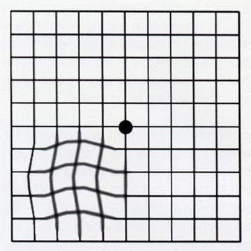
Amsler grid with wavy lines
Following are some common ways vision loss is detected:
- words on a page look blurred
- a dark or empty area appears in the center of vision
- straight lines look distorted, as in the diagram on the right shows
How is macular degeneration diagnosed?
Many people do not realize that they have a macular problem until blurred vision becomes obvious. Your ophthalmologist (Eye M.D.) can detect early stages of AMD during a medical eye examination that includes the following:
- a simple vision test in which you look at a chart that resembles graph paper (Amsler grid)
- viewing the macula with an ophthalmoscope
- taking special photographs of the eye called OCT or Optical Coherence Tomography and fluorescein angiography to find abnormal blood vessels under the retina
Wet AMD
The Macula and How the Eye Sees
Dry AMD
Retinal Angiography
- Anatomy of the Eye
- Botox
- Cataracts
- Diabetes and the Eye
- Diabetic Retinopathy – What is it and how is it detected?
- Treatment for Diabetic Retinopathy
- Non-Proliferative Diabetic Retinopathy (NPDR) – Video
- Proliferative Diabetic Retinopathy (PDR) – Video
- Cystoid Macular Edema
- Vitreous Hemorrhage – Bleeding from diabetes (Video)
- Vitrectomy Surgery for Vitreous Hemorrhage (Video)
- Macular Edema
- Laser Procedures for Macular Edema (Video)
- Laser for Proliferative Diabetic Retinopathy – PDR (Video)
- How the Eye Sees (Video)
- Dilating Eye Drops
- Dry Eyes and Tearing
- Eye Lid Problems
- A Word About Eyelid Problems
- Bells Palsy
- Blepharitis
- Blepharoptosis – Droopy Eyelids (Video)
- Dermatochalasis – excessive upper eyelid skin (Video)
- Ectropion – Sagging Lower Eyelids (Video)
- Entropion – Inward Turning Eyelids (Video)
- How to Apply Warm Compresses
- Ocular Rosacea
- Removing Eyelid Lesions
- Styes and Chalazion
- Twitches or Spasms
- Floaters and Flashes
- Glaucoma
- Selective Laser Trabeculoplasty (SLT) for Glaucoma
- Glaucoma: What is it and how is it detected?
- Optical Coherence Tomography OCT – Retina & Optic Nerve Scan
- Treatment for Glaucoma
- Retinal Nerve Fibers and Glaucoma (Video)
- Open Angle Glaucoma (Video)
- Closed Angle Glaucoma (Video)
- Visual Field Test for Glaucoma
- Glaucoma and Blind Spots (Video)
- Treatment for Glaucoma with Laser Iridotomy (Video)
- Laser Treatment for Glaucoma with ALT and SLT (Video)
- Surgical Treatment for Glaucoma with Trabeculectomy (Video)
- Surgical Treatment of Glaucoma with Seton (Video)
- Keeping Eyes Healthy
- Laser Vision Correction
- Latisse for Eyelashes
- Macular Degeneration
- Macular Degeneration – What is it and how is it detected?
- Treatment for Macular Degeneration
- Dry Macular Degeneration (Video)
- Wet Macular Degeneration (Video)
- Treatment of Macular Degeneration with Supplements
- Treatment of Wet Macular Degeneration with Anti-VEGF Injections
- Amsler Grid – A home test for Macular Degeneration (Video)
- Living with Vision Loss
- How the Eye Works – The Macula (Video)
- Other Eye Conditions
- Central Serous Retinopathy
- Lattice Degeneration of the Retina
- A Word About Other Eye Conditions
- Amblyopia
- Carotid Artery Disease and the Eye
- Fuch’s Corneal Dystrophy
- Herpes Simplex and the Eye
- Herpes Zoster (Shingles) and the Eye
- Ischemic Optic Neuropathy
- Keratoconus
- Macular Hole
- Macular Pucker
- Microvascular Cranial Nerve Palsy
- Migraine and the Eye
- Optic Neuritis
- Pseudotumor Cerebri
- Retinal Vein Occlusion
- Retinitis Pigmentosa
- Retinopathy of Prematurity
- Strabismus
- Thyroid Disorders and the Eye
- Uveitis
- Vitreomacular Adhesions / Vitreomacular Traction Syndrome
- Red Eye
- Refractive Errors
- Retinal Tears and Detachments
Disclaimer
This Patient Education Center is provided for informational and educational purposes only. It is NOT intended to provide, nor should you use it for, instruction on medical diagnosis or treatment, and it does not provide medical advice. The information contained in the Patient Education Center is compiled from a variety of sources. It does NOT cover all medical problems, eye diseases, eye conditions, ailments or treatments.
You should NOT rely on this information to determine a diagnosis or course of treatment. The information should NOT be used in place of an individual consultation, examination, visit or call with your physician or other qualified health care provider. You should never disregard the advice of your physician or other qualified health care provider because of any information you read on this site or any web sites you visit as a result of this site.
Promptly consult your physician or other qualified health provider if you have any health care questions or concerns and before you begin or alter any treatment plan. No doctor-patient relationship is established by your use of this site.


