Overflow Tearing and Chronic Infections in Infants
Abnormal or overflow eye tearing is a common condition in infants.
In fact, approximately one-third of all newborns have excessive tears and mucus. It occurs when a membrane (a skin-like tissue) in the nose fails to open before birth, blocking part of the tear drainage system. If tears do not drain properly, they can collect inside the tear drainage system and spill over the eyelid onto the cheek. The tear sac can also become infected, which may lead to the development of conjunctivitis, commonly known as pink eye. You should contact your primary care physician or ophthalmologist (Eye M.D.) if the discharge becomes thicker or changes color from white to yellow or green, or the white of the eye becomes red.

The eye’s tear drainage system
How do tears drain from the eye?
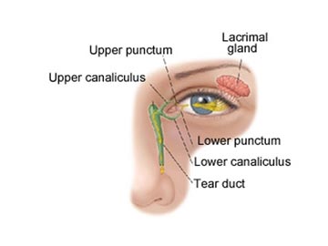
Tears are produced to keep your eyes moist. As new tears are produced, old tears drain from the eye through two small holes called the upper and lower puncta, located at the corner of your upper and lower eyelids near the nose. The tears then move through a passage called the canaliculus and into the lacrimal sac. From the sac, the tears drop down the tear duct (called nasolacrimal duct), which drains into the back of your nose and throat. That is why your nose runs when you cry.
See this short video on The Tear Drainage System.
In infants with tear duct obstruction, there is a membrane at the end (bottom) of the tear duct preventing tears from draining into the back of the nose and throat.
Are there other causes of tearing?
A very rare condition called congenital glaucoma can also cause excessive tearing. With congenital glaucoma, other signs and symptoms will accompany tearing, such as an enlarged eye, a cloudy cornea, high eye pressure, light sensitivity and eye irritation.
Tearing can also be caused by wind, smoke, allergies or other environmental irritants. Rarely, the tear drainage system fails to develop normally. An eye examination by an ophthalmologist will identify the exact cause of the tearing.
How is overflow tearing treated?
Your ophthalmologist may recommend:
- applying antibiotic eyedrops or ointment to the eye once or twice daily to fight infection
- cleaning the eyelids with warm water
- applying pressure (or massage) over the lacrimal sac.
The purpose of massage is to put pressure on the lacrimal sac to pop open the membrane at the bottom of the tear duct. This is most easily accomplished by placing your hands on each side of the baby’s face with your index finger(s) between the inner corner of the eye and the side of the nose, pressing in and down over the lacrimal sac for a few seconds. This should be done several times a day, such as at each diaper change.
The blocked tear duct often spontaneously opens within six to 12 months after birth. If overflow tearing persists, it may be necessary for your ophthalmologist to open the obstruction surgically by passing a probe through the tear duct.
How is probing of the tear duct performed?
A thin, metal probe is gently inserted through the tear drainage system to open the obstruction. The drainage system is then flushed with fluid to make sure the pathway is open. The procedure is performed in an outpatient setting under local or general anesthesia. It causes little or no pain, but tears may be stained briefly with blood or a nosebleed may occur. An antibiotic eye drop may be prescribed.
Are any risks involved with probing?
As with any surgical procedure, complications can occur, including:
- infection
- bleeding
- re-obstruction of the tear duct
Re-obstruction of the tear duct may require another probing or additional surgery. Be sure to discuss potential complications with your ophthalmologist before surgery.
- Anatomy of the Eye
- Botox
- Cataracts
- Diabetes and the Eye
- Diabetic Retinopathy – What is it and how is it detected?
- Treatment for Diabetic Retinopathy
- Non-Proliferative Diabetic Retinopathy (NPDR) – Video
- Proliferative Diabetic Retinopathy (PDR) – Video
- Cystoid Macular Edema
- Vitreous Hemorrhage – Bleeding from diabetes (Video)
- Vitrectomy Surgery for Vitreous Hemorrhage (Video)
- Macular Edema
- Laser Procedures for Macular Edema (Video)
- Laser for Proliferative Diabetic Retinopathy – PDR (Video)
- How the Eye Sees (Video)
- Dilating Eye Drops
- Dry Eyes and Tearing
- Eye Lid Problems
- A Word About Eyelid Problems
- Bells Palsy
- Blepharitis
- Blepharoptosis – Droopy Eyelids (Video)
- Dermatochalasis – excessive upper eyelid skin (Video)
- Ectropion – Sagging Lower Eyelids (Video)
- Entropion – Inward Turning Eyelids (Video)
- How to Apply Warm Compresses
- Ocular Rosacea
- Removing Eyelid Lesions
- Styes and Chalazion
- Twitches or Spasms
- Floaters and Flashes
- Glaucoma
- Selective Laser Trabeculoplasty (SLT) for Glaucoma
- Glaucoma: What is it and how is it detected?
- Optical Coherence Tomography OCT – Retina & Optic Nerve Scan
- Treatment for Glaucoma
- Retinal Nerve Fibers and Glaucoma (Video)
- Open Angle Glaucoma (Video)
- Closed Angle Glaucoma (Video)
- Visual Field Test for Glaucoma
- Glaucoma and Blind Spots (Video)
- Treatment for Glaucoma with Laser Iridotomy (Video)
- Laser Treatment for Glaucoma with ALT and SLT (Video)
- Surgical Treatment for Glaucoma with Trabeculectomy (Video)
- Surgical Treatment of Glaucoma with Seton (Video)
- Keeping Eyes Healthy
- Laser Vision Correction
- Latisse for Eyelashes
- Macular Degeneration
- Macular Degeneration – What is it and how is it detected?
- Treatment for Macular Degeneration
- Dry Macular Degeneration (Video)
- Wet Macular Degeneration (Video)
- Treatment of Macular Degeneration with Supplements
- Treatment of Wet Macular Degeneration with Anti-VEGF Injections
- Amsler Grid – A home test for Macular Degeneration (Video)
- Living with Vision Loss
- How the Eye Works – The Macula (Video)
- Other Eye Conditions
- Central Serous Retinopathy
- Lattice Degeneration of the Retina
- A Word About Other Eye Conditions
- Amblyopia
- Carotid Artery Disease and the Eye
- Fuch’s Corneal Dystrophy
- Herpes Simplex and the Eye
- Herpes Zoster (Shingles) and the Eye
- Ischemic Optic Neuropathy
- Keratoconus
- Macular Hole
- Macular Pucker
- Microvascular Cranial Nerve Palsy
- Migraine and the Eye
- Optic Neuritis
- Pseudotumor Cerebri
- Retinal Vein Occlusion
- Retinitis Pigmentosa
- Retinopathy of Prematurity
- Strabismus
- Thyroid Disorders and the Eye
- Uveitis
- Vitreomacular Adhesions / Vitreomacular Traction Syndrome
- Red Eye
- Refractive Errors
- Retinal Tears and Detachments
Disclaimer
This Patient Education Center is provided for informational and educational purposes only. It is NOT intended to provide, nor should you use it for, instruction on medical diagnosis or treatment, and it does not provide medical advice. The information contained in the Patient Education Center is compiled from a variety of sources. It does NOT cover all medical problems, eye diseases, eye conditions, ailments or treatments.
You should NOT rely on this information to determine a diagnosis or course of treatment. The information should NOT be used in place of an individual consultation, examination, visit or call with your physician or other qualified health care provider. You should never disregard the advice of your physician or other qualified health care provider because of any information you read on this site or any web sites you visit as a result of this site.
Promptly consult your physician or other qualified health provider if you have any health care questions or concerns and before you begin or alter any treatment plan. No doctor-patient relationship is established by your use of this site.


