Pseudotumor Cerebri
Pseudotumor cerebri (PTC) is a condition in which high cerebrospinal fluid (CSF) pressure inside your head can cause problems with vision and headache.
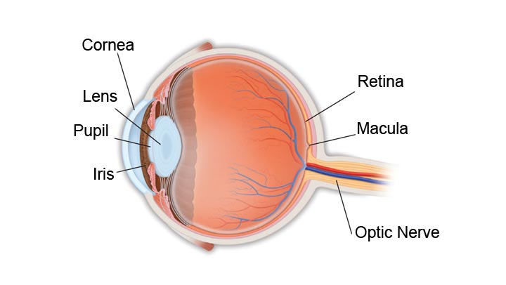
The term “pseudotumor” (which means “false tumor”) comes from the days before CT and MRI scans, when doctors who noted swelling of the optic disc (the visible portion of the optic nerve in the back of the eye) considered the possibility of a brain tumor.

Patients with optic disc swelling but no evidence of a tumor were said to have “pseudotumor.” In PTC, the flow of CSF (a clear fluid that bathes the brain and spinal cord) is blocked from flowing back out of the head as it should, leading to high CSF pressure inside your head. This pressure results in swelling of the optic disc at the back of the eye, which can damage (sometimes permanently) the optic nerve and cause vision loss. High pressure may also cause damage to the nerves that move the eyes, resulting in double vision.
What causes pseudotumor cerebri?
The reason for decreased outflow of CSF is not clear. Because this condition seems to occur more often in overweight young women, a hormonal influence is suspected.
Overweight young women are 20 times more likely to develop pseudotumor. PTC may also occur in children, men, and patients who are not overweight. In some cases, antibiotic or steroid use may be associated with pseudotumor. High doses of vitamin A may also lead to increased CSF pressure.
What are the symptoms of pseudotumor cerebri?
The most common symptoms of high CSF pressure inside the head are headache and visual loss. The headache may be located anywhere but is frequently in the back of the neck. This pain may awaken the patient in the middle of the night and may worsen with bending or stooping. Symptoms of pseudotumor cerebri include:
- Dimming, blurring or graying of vision
- Difficulty seeing to the side
- Brief visual disturbances (often associated with bending or stooping)
- Double vision
- Rushing noise in the ears
- Nausea and vomiting
How is pseudotumor cerebri diagnosed?
Your ophthalmologist (Eye M.D.) will carefully measure your vision, check the light reaction of your pupils, examine the back of your eye and may evaluate your field of vision. Because other conditions may produce similar symptoms to pseudotumor cerebri, an MRI scan is necessary for accurate diagnosis. A spinal tap is also necessary to check for elevated CSF and to make sure there are no other CSF abnormalities.
How is pseudotumor cerebri treated?
If you have no significant headaches or evidence of vision loss (including visual fields), no treatment may be necessary. If you do experience these problems, however, certain medications used in treating glaucoma (such as acetazolamide) can lower the CSF pressure in the head by reducing production of this fluid. Diuretics may also be prescribed. One of the most effective treatments is weight reduction in overweight patients. Pressure may also be lowered by draining off CSF through repeated spinal taps.
Repeat visual field testing is essential in following patients with PTC. If your visual field is worsening or you experience a decrease in central vision, and you do not have severe headaches, a small hole or multiple slits may be placed in the optic nerve sheath (called optic nerve sheath fenestration) just behind the eye using an operating microscope. This is done to protect the optic nerve from further damage. If severe headaches accompany visual loss, a shunting procedure (lumbo-peritoneal or ventriculo-peritoneal) may be required, in which a small tube is placed to carry fluid from where it is building up to where it can be absorbed, thus relieving pressure.
- Anatomy of the Eye
- Botox
- Cataracts
- Diabetes and the Eye
- Diabetic Retinopathy – What is it and how is it detected?
- Treatment for Diabetic Retinopathy
- Non-Proliferative Diabetic Retinopathy (NPDR) – Video
- Proliferative Diabetic Retinopathy (PDR) – Video
- Cystoid Macular Edema
- Vitreous Hemorrhage – Bleeding from diabetes (Video)
- Vitrectomy Surgery for Vitreous Hemorrhage (Video)
- Macular Edema
- Laser Procedures for Macular Edema (Video)
- Laser for Proliferative Diabetic Retinopathy – PDR (Video)
- How the Eye Sees (Video)
- Dilating Eye Drops
- Dry Eyes and Tearing
- Eye Lid Problems
- A Word About Eyelid Problems
- Bells Palsy
- Blepharitis
- Blepharoptosis – Droopy Eyelids (Video)
- Dermatochalasis – excessive upper eyelid skin (Video)
- Ectropion – Sagging Lower Eyelids (Video)
- Entropion – Inward Turning Eyelids (Video)
- How to Apply Warm Compresses
- Ocular Rosacea
- Removing Eyelid Lesions
- Styes and Chalazion
- Twitches or Spasms
- Floaters and Flashes
- Glaucoma
- Selective Laser Trabeculoplasty (SLT) for Glaucoma
- Glaucoma: What is it and how is it detected?
- Optical Coherence Tomography OCT – Retina & Optic Nerve Scan
- Treatment for Glaucoma
- Retinal Nerve Fibers and Glaucoma (Video)
- Open Angle Glaucoma (Video)
- Closed Angle Glaucoma (Video)
- Visual Field Test for Glaucoma
- Glaucoma and Blind Spots (Video)
- Treatment for Glaucoma with Laser Iridotomy (Video)
- Laser Treatment for Glaucoma with ALT and SLT (Video)
- Surgical Treatment for Glaucoma with Trabeculectomy (Video)
- Surgical Treatment of Glaucoma with Seton (Video)
- Keeping Eyes Healthy
- Laser Vision Correction
- Latisse for Eyelashes
- Macular Degeneration
- Macular Degeneration – What is it and how is it detected?
- Treatment for Macular Degeneration
- Dry Macular Degeneration (Video)
- Wet Macular Degeneration (Video)
- Treatment of Macular Degeneration with Supplements
- Treatment of Wet Macular Degeneration with Anti-VEGF Injections
- Amsler Grid – A home test for Macular Degeneration (Video)
- Living with Vision Loss
- How the Eye Works – The Macula (Video)
- Other Eye Conditions
- Central Serous Retinopathy
- Lattice Degeneration of the Retina
- A Word About Other Eye Conditions
- Amblyopia
- Carotid Artery Disease and the Eye
- Fuch’s Corneal Dystrophy
- Herpes Simplex and the Eye
- Herpes Zoster (Shingles) and the Eye
- Ischemic Optic Neuropathy
- Keratoconus
- Macular Hole
- Macular Pucker
- Microvascular Cranial Nerve Palsy
- Migraine and the Eye
- Optic Neuritis
- Pseudotumor Cerebri
- Retinal Vein Occlusion
- Retinitis Pigmentosa
- Retinopathy of Prematurity
- Strabismus
- Thyroid Disorders and the Eye
- Uveitis
- Vitreomacular Adhesions / Vitreomacular Traction Syndrome
- Red Eye
- Refractive Errors
- Retinal Tears and Detachments
Disclaimer
This Patient Education Center is provided for informational and educational purposes only. It is NOT intended to provide, nor should you use it for, instruction on medical diagnosis or treatment, and it does not provide medical advice. The information contained in the Patient Education Center is compiled from a variety of sources. It does NOT cover all medical problems, eye diseases, eye conditions, ailments or treatments.
You should NOT rely on this information to determine a diagnosis or course of treatment. The information should NOT be used in place of an individual consultation, examination, visit or call with your physician or other qualified health care provider. You should never disregard the advice of your physician or other qualified health care provider because of any information you read on this site or any web sites you visit as a result of this site.
Promptly consult your physician or other qualified health provider if you have any health care questions or concerns and before you begin or alter any treatment plan. No doctor-patient relationship is established by your use of this site.


