Treatment for Glaucoma with Laser Iridotomy (Video)
Laser iridotomy is a surgical procedure used to treat angle closure glaucoma and those at risk for angle closure glaucoma.
As with many medical conditions, it is preferable to treat patients at risk and thereby avoid vision loss.
What is angle-closure glaucoma?
Like other forms of glaucoma, angle-closure glaucoma has to do with pressure inside the eye. A normal eye constantly produces a certain amount of clear liquid called aqueous , which circulates inside the front portion of the eye. An equal amount of this fluid flows out of the eye through a very tiny drainage system called the drainage angle, thus maintaining a constant level of pressure within the eye.
There are two main types of glaucoma. The most common type is open-angle glaucoma, in which fluid drains too slowly from the eye and causes a chronic rise in eye pressure. In contrast, angle-closure glaucoma causes a more sudden rise in eye pressure. In angle-closure glaucoma, the drainage angle may become partially or completely blocked when the iris (the colored part of the eye) is pushed over this area. The iris may completely block the aqueous fluid from leaving the eye, much like a stopper in a sink. In this situation, the pressure inside the eye can rise very quickly and cause an acute angle-closure glaucoma attack.
Symptoms of an acute angle-closure glaucoma attack include severe ocular pain and redness, decreased vision, colored halos, headache, nausea and vomiting. Because raised eye pressure can rapidly damage the optic nerve and lead to vision loss, an angle-closure glaucoma attack must be treated immediately.
Unfortunately, individuals at risk of developing angle closure glaucoma often have few or no symptoms prior to the attack. Risk factors for angle-closure glaucoma include increasing age, farsightedness (hyperopia), and Asian heritage. Some early symptoms in people at risk for angle-closure glaucoma include blurred vision, halos in their vision, headache, mild eye pain or redness.
People who are at risk for developing angle-closure glaucoma should have a laser iridotomy. Many common medications, including over-the-counter cold medications and sleeping pills (and any other medication that can dilate the pupil), should be avoided until after the laser procedure is completed. If one eye has an attack of angle-closure glaucoma, the other eye is also at risk and may need treatment.
What happens during laser iridotomy?

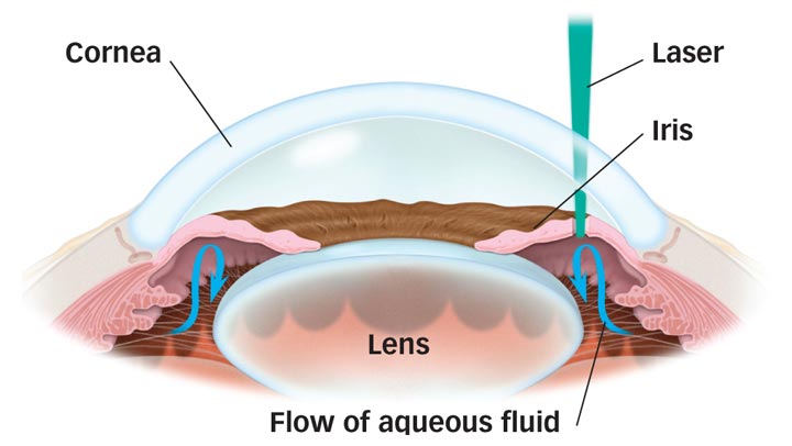
A laser is used to make a small hole in the iris.

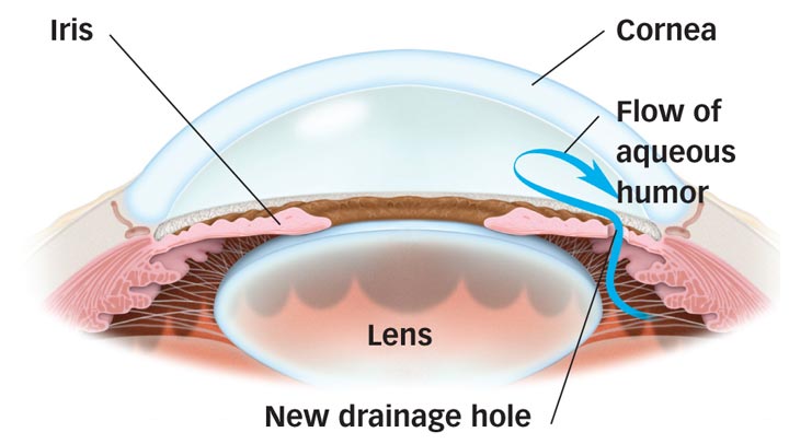
After the laser treatment, aqueous fluid flows through the new iridotomy, reducing the risk of acute closed angle glaucoma.
Using a laser, a small hole is made in the iris to create a new pathway for the aqueous fluid to drain from your eye. The new drainage hole allows the iris to fall back into its normal position, restoring the balance between fluid entering and leaving your eye and lowering the eye pressure.
The surgery is performed by your ophthalmologist (Eye M.D.) on an outpatient basis, usually in his or her office. Your eye will be numbed with eyedrops.
After the laser treatment, aqueous fluid flows through the new iridotomy, reducing the risk of acute closed angle glaucoma.
A contact lens is placed on your eye to serve as a precise guide for the laser. A hole about the size of a pinhead is made in your iris, and will be concealed from view by your upper eyelid. The actual procedure will only take a few minutes. You should plan to have someone drive you home afterward.
Are there any risks or side effects?
Complications following laser iridotomy are uncommon. They include:
- A spike in eye pressure
- Inflammation
- Cataract
- Bleeding
- Need for re-treatment
- Blurred vision
- Light image or streak
- Pain
The risks and side effects of glaucoma treatment are always balanced with the greater risk of leaving glaucoma untreated.
Laser Iridotomy
Article Videos
- Anatomy of the Eye
- Botox
- Cataracts
- Diabetes and the Eye
- Diabetic Retinopathy – What is it and how is it detected?
- Treatment for Diabetic Retinopathy
- Non-Proliferative Diabetic Retinopathy (NPDR) – Video
- Proliferative Diabetic Retinopathy (PDR) – Video
- Cystoid Macular Edema
- Vitreous Hemorrhage – Bleeding from diabetes (Video)
- Vitrectomy Surgery for Vitreous Hemorrhage (Video)
- Macular Edema
- Laser Procedures for Macular Edema (Video)
- Laser for Proliferative Diabetic Retinopathy – PDR (Video)
- How the Eye Sees (Video)
- Dilating Eye Drops
- Dry Eyes and Tearing
- Eye Lid Problems
- A Word About Eyelid Problems
- Bells Palsy
- Blepharitis
- Blepharoptosis – Droopy Eyelids (Video)
- Dermatochalasis – excessive upper eyelid skin (Video)
- Ectropion – Sagging Lower Eyelids (Video)
- Entropion – Inward Turning Eyelids (Video)
- How to Apply Warm Compresses
- Ocular Rosacea
- Removing Eyelid Lesions
- Styes and Chalazion
- Twitches or Spasms
- Floaters and Flashes
- Glaucoma
- Selective Laser Trabeculoplasty (SLT) for Glaucoma
- Glaucoma: What is it and how is it detected?
- Optical Coherence Tomography OCT – Retina & Optic Nerve Scan
- Treatment for Glaucoma
- Retinal Nerve Fibers and Glaucoma (Video)
- Open Angle Glaucoma (Video)
- Closed Angle Glaucoma (Video)
- Visual Field Test for Glaucoma
- Glaucoma and Blind Spots (Video)
- Treatment for Glaucoma with Laser Iridotomy (Video)
- Laser Treatment for Glaucoma with ALT and SLT (Video)
- Surgical Treatment for Glaucoma with Trabeculectomy (Video)
- Surgical Treatment of Glaucoma with Seton (Video)
- Keeping Eyes Healthy
- Laser Vision Correction
- Latisse for Eyelashes
- Macular Degeneration
- Macular Degeneration – What is it and how is it detected?
- Treatment for Macular Degeneration
- Dry Macular Degeneration (Video)
- Wet Macular Degeneration (Video)
- Treatment of Macular Degeneration with Supplements
- Treatment of Wet Macular Degeneration with Anti-VEGF Injections
- Amsler Grid – A home test for Macular Degeneration (Video)
- Living with Vision Loss
- How the Eye Works – The Macula (Video)
- Other Eye Conditions
- Central Serous Retinopathy
- Lattice Degeneration of the Retina
- A Word About Other Eye Conditions
- Amblyopia
- Carotid Artery Disease and the Eye
- Fuch’s Corneal Dystrophy
- Herpes Simplex and the Eye
- Herpes Zoster (Shingles) and the Eye
- Ischemic Optic Neuropathy
- Keratoconus
- Macular Hole
- Macular Pucker
- Microvascular Cranial Nerve Palsy
- Migraine and the Eye
- Optic Neuritis
- Pseudotumor Cerebri
- Retinal Vein Occlusion
- Retinitis Pigmentosa
- Retinopathy of Prematurity
- Strabismus
- Thyroid Disorders and the Eye
- Uveitis
- Vitreomacular Adhesions / Vitreomacular Traction Syndrome
- Red Eye
- Refractive Errors
- Retinal Tears and Detachments
Disclaimer
This Patient Education Center is provided for informational and educational purposes only. It is NOT intended to provide, nor should you use it for, instruction on medical diagnosis or treatment, and it does not provide medical advice. The information contained in the Patient Education Center is compiled from a variety of sources. It does NOT cover all medical problems, eye diseases, eye conditions, ailments or treatments.
You should NOT rely on this information to determine a diagnosis or course of treatment. The information should NOT be used in place of an individual consultation, examination, visit or call with your physician or other qualified health care provider. You should never disregard the advice of your physician or other qualified health care provider because of any information you read on this site or any web sites you visit as a result of this site.
Promptly consult your physician or other qualified health provider if you have any health care questions or concerns and before you begin or alter any treatment plan. No doctor-patient relationship is established by your use of this site.


